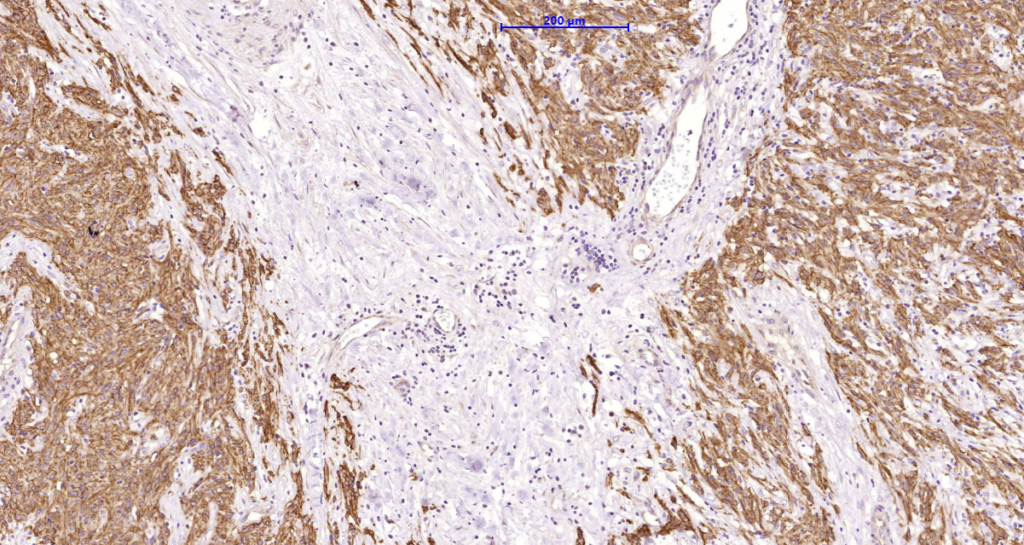
Expression of DOG-1 protein is elevated in the gastrointestinal stromal tumors (GIST’s), KIT signaling-driven mesenchymal tumors of the GI tract. DOG-1 is rarely expressed in other soft tissue tumors, which, due to appearance, may be difficult to diagnose. Immunoreactivity for DOG-1 has been reported in 97.8 percent of scorable GIST’s, including all KIT negative GIST’s. Overexpression of DOG-1 has been suggested to aid in the identification of GISTs, including Platelet-Derived Growth Factor Receptor Alpha mutants that fail to express KIT antigen. The overall sensitivity of DOG1 and KIT in GIST’s is nearly identical: 94.4% vs. 94.7%.
Availability:
| Catalog No. | Contents | Volume |
| ILM1484-C01 | DOG-1 | 0,1 ml concentrate |
| ILM1484-C05 | DOG-1 | 0,5 ml concentrate |
| ILM1484-C1 | DOG-1 | 1,0 ml concentrate |
Reactivity: Human
Clone: DG/1447
Species of origin: Mouse
Isotype: IgG1 k
Control Tissue: Gastrointestinal Stromal Tumor (GIST) or testicular germ cell tumor. Melanocytes in the basal layer of the epidermis and mast cells in the dermis of normal skin.
Staining: Cytoplasmic and membranous
Immunogen: Recombinant human DOG-1 protein
Presentation: Bioreactor Concentrate with 0.05% Azide
Application and suggested dilutions:
Pretreatment: Heat induced epitope retrieval in 10 mM citrate buffer, pH6.0, or in 50 mM Tris buffer pH9.5, for 20 minutes is required for IHC staining on formalin-fixed, paraffin embedded tissue sections.
- Immunohistochemical staining of formalin-fixed, paraffin embedded tissue section (dilution 1:200-1:400)
- Immunofluorescence dilution up to 1:50-1:100
- Flow Cytometry 5µl per test per one millions cells or 5µl per 100µl of whole blood.
The optimal dilution for a specific application should be determined by the investigator.

