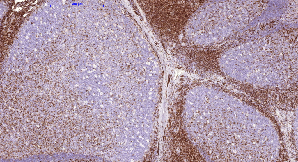
It recognizes a cell surface glycoprotein of 95/115/135kDa (depending upon the extent of glycosylation), identified as CD43. 70-90% if T-cell lymphomas and from 22-37% of B-cell lymphomas express CD43. No reactivity has been observed with reactive B-cells. So a B-lineage population that co-expresses CD43 is highly likely to be malignant lymphoma, especially a low grade lymphoma, rather than a reactive B-cell population. When CD43 antibody is used in combination with anti-CD20, effective immunophenotyping of the lymphomas in formalin-fixed tissues can be obtained. Co-staining of a lymphoid infiltrate with anti-CD20 and anti-CD43 argues against a reactive process and favors a diagnosis of lymphoma.
Availability:
| Catalog No. | Contents | Volume |
| ILM3388-C01 | CD43 | 0.1 ml concentrate |
| ILM3388-C05 | CD43 | 0,5 ml concentrate |
| ILM3388-C1 | CD43 | 1.0 ml concentrate |
Reactivity: Human
Clone: SPN/3388
Human Entrez Gene ID: 6693
Human SwissProt: P16150
Human Unigene: 632188
Species of origin: Mouse
Isotype: IgG1, k
Control Tissue: Human mantle cell lymphoma tissue
Staining: Cell surface
Immunogen: Recombinant full-length human SPN protein
Presentation:
Purified antibody from bioreactor concentrate by protein A/G. Prepared in 10mM PBS with 0.05% BSA and 0.05% Azide.
Application and suggested dilutions:
Pretreatment: Heat induced epitope retrieval in 10 mM citrate buffer, pH6.0, for 20 minutes is required for IHC staining on formalin-fixed, paraffin embedded tissue sections.
- Immunohistochemical staining of cryostat tissue sections (dilution up to 1:100-1:200)
- Immunohistochemical staining of formalin-fixed, paraffin embedded tissue section
(dilution 1:100 to 1:200). - Western blot (dilution 1:100 – 1:200)
The optimal dilution for a specific application should be determined by the investigator.

