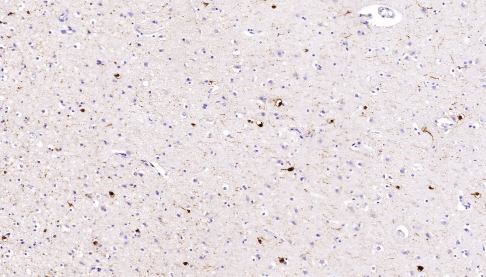
Mouse Monoclonal Antibody
It recognizes a protein of about 29kDa, which is identified as Calretinin (also known as Calbindin 2). Calretinin is a vitamin D-dependent calcium-binding protein involved in calcium signaling. It is present in subsets of neurons throughout the brain and spinal chord, including sensory ganglia. Antibody to calretinin is useful in differentiating mesothelioma from adenocarcinomas of the lung. It also aids in differentiating adrenal cortical neoplasms from pheochromocytomas.
Availability:
| Catalog No. | Contents | Volume |
| ILM2786-C01 | Calretinin | 0,1 ml concentrate |
| ILM2786-C05 | Calretinin | 0,5 ml concentrate |
| ILM2786-C1 | Calretinin | 1,0 ml concentrate |
Intended use: For Research Use Only
Reactivity: Human, others not known
Clone: CALB/2786
Human Gene ID: 794
Human SwissProt: P22676
Human Unigene: 106857
Species of origin: Mouse
Isotype: IgG1, Kappa
Immunogen: Recombinant human Calretinin (Calbindin 2) protein fragment (around aa23-243) (exact sequence is proprietary)
Control Tissue: Brain, Testis or Mesothelioma
Staining: nuclear and cytoplasmatic
Presentation: Bioreactor concentrate with 0.05% Azide
Application and suggested dilutions:
Pre-treatment: Heat induced epitope retrieval in 10 mM citrate buffer pH6.0 for 20 minutes is required for IHC staining on formalin-fixed, paraffin embedded tissue sections.
- Immunohistochemical staining of formalin-fixed, paraffin embedded tissue section (dilution up to 1:100)
- Western blot (1-2ug/ml)
The optimal dilution for a specific application should be determined by the investigator.
Note: Dilution of the antibody in 10% normal goat serum followed by a Goat anti-Mouse secondary antibody-based detection is recommended.
Storage & Stability: Store at 2-8 °C. Do not use after expiration date printed on the vial.
Reference:
- Doglioni, C., et al. 1996. AM J. Surg. Pathol. 20:1037-1046
- Rogers, J.H. 1987. J. Cell Biol. 105:1343-1353

