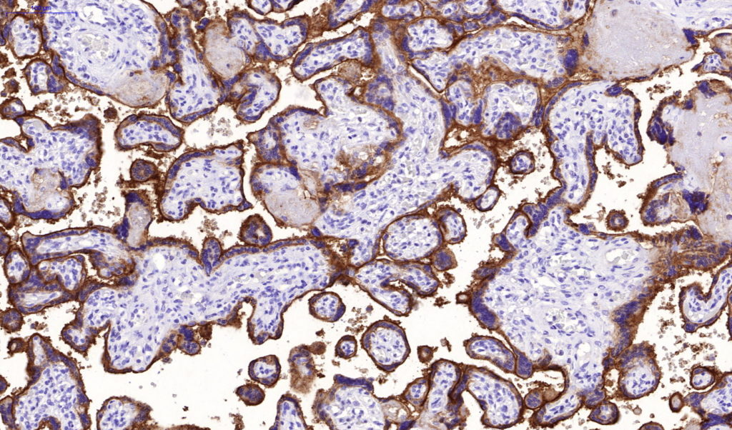
Recombinant Mouse Monoclonal Antibody
PD-L1 is a checkpoint regulator in immune cells, it is expressed on immune or non-hematopoietic cells. Expression of the protein is seen during pregnancy where it has a role in suppressing the immune system. PD-L1 induces an inhibitory signal in activated T-cells and promotes T-cell apoptosis. It is overexpressed in a number of different cancers where it is believed to play a significant role in the cancer s ability to evade the immune system.
Availability:
| Catalog No. | Contents | Volume |
| ILM2740-C01 | CD274 | 0,1 ml concentrate |
| ILM2740-C05 | CD274 | 0,5 ml concentrate |
| ILM2740-C1 | CD274 | 1,0 ml concentrate |
Intended use: For Research Use Only
Reactivity: Human
Clone: rPDL1/4772
Species of origin: Recombinant Mouse
Isotype: IgG2a, Kappa
Gene ID: 29126
SwissProt: Q9NZQ7
Unigene: 521989
Control Tissue: Placenta, spleen, tonsil
Staining: Membranous and cytoplasmatic
Immunogen: KLH-conjugated linear peptide corresponding to human PD-L1
Presentation: Bioreactor Concentrate with 0.05% Azide
Application and suggested dilutions:
Pretreatment: Heat induced epitope retrieval in 10 mM Citrate buffer pH6.0 for 20 minutes is required for IHC staining on formalin-fixed, paraffin embedded tissue sections.
- Immunohistochemical staining of formalin-fixed, paraffin embedded tissue section (dilution 1:50-1:100)
The optimal dilution for a specific application should be determined by the investigator.
Note: Dilution of the antibody in 10% normal goat serum followed by a Goat anti-Mouse secondary antibody-based detection is recommended.
Storage & Stability: Store at 2-8 °C. Do not use after expiration date printed on the vial.

