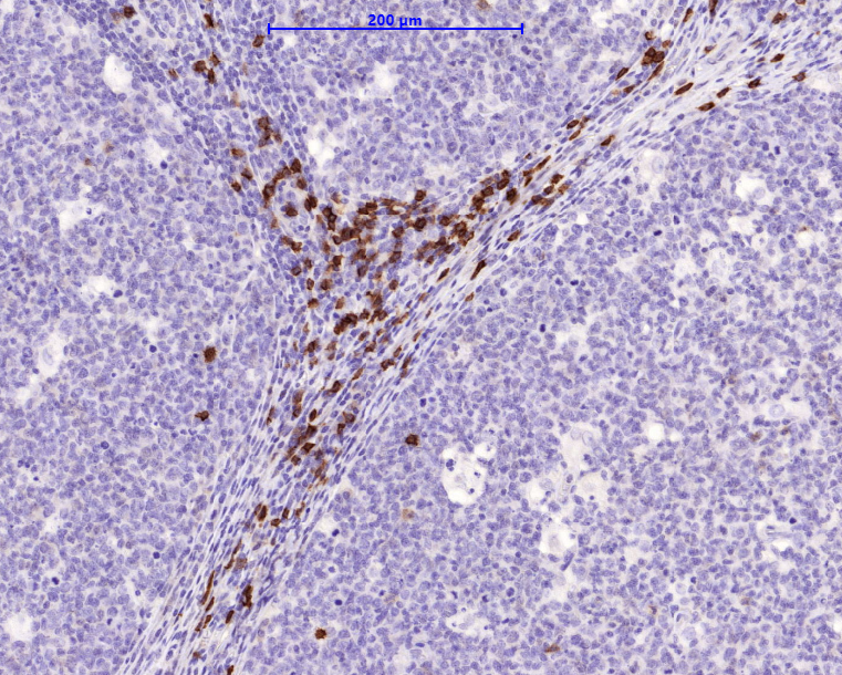
CD8a is a cell surface receptor expressed either as a heterodimer with the CD8 beta chain (CD8 alpha/beta) or as a homodimer (CD8 alpha/alpha). A majority of thymocytes and a subpopulation of mature T cells and NK cells express CD8a. CD8 binds to MHC class 1 and trough its association with protein tyrosine kinase p56lck plays a role in T cell development and activation of mature T cells. For mature T-cells, CD4 and CD8 are mutually exclusive, so anti-CD8 generally used in conjunction with anti-CD4. It is a useful marker for distinguishing helper/inducer T-Lymphocytes and most peripheral T-cell lymphomas are CD4+/CD8-. Anaplastic large cells lymphoma is usually CD4+ and CD8-, and in T-lymphoblastic lymphoma/leukemia, CD4 and CD8 are often co-expressed. CD8 is also found in littoral cell angioma of the spleen.
Availability:
| Catalog no. | Contents | Volume |
| ILM9250-C01 | CD8a | 0,1 ml concentrate |
| ILM9250-C05 | CD8a | 0,5 ml concentrate |
| ILM9250-C1 | CD8a | 1,0 ml concentrate |
Reactivity: Human
Clone: C8/1779R
Species of origin: Rabbit
Isotype: IgG
Control Tissue: HuT78 or hPBL, Tonsil
Staining: Cell Surface
Immunogen: Recombinant Full-length human CD8a protein
Presentation: Bioreactor Concentrate with 0.05% Azide
Application and suggested dilutions:
Pretreatment: Heat induced epitope retrieval in 10 mM citrate buffer, pH6.0 for 20 minutes is required for IHC staining on formalin-fixed, paraffin embedded tissue sections.
- Immunohistochemical staining of cryostat tissue sections (dilution up to 1:100-1:200)
- Immunohistochemical staining of formalin-fixed, paraffin embedded tissue section (dilution up to 1:100-1:200)
The optimal dilution for a specific application should be determined by the investigator.

